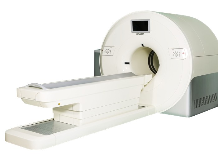
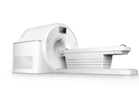
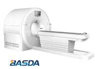
Virtually no deformation is occurring with the Detector Channels during rotation due to the rigidity of the rotor. The stability of the detector subsystem allows for stable, consistent and accurate image generation.
System Software
Conduct random correction, scattering correction, attenuation correction, and uniformity correction.
- Beam hardening artifact correction software
Beam hardening correction technology is able to reduce the artifacts in the transition region between the bone and the soft tissue. It is also suitable for pelvis scanning.
- Posterior cranial fossa optimization
Equipped with the abundant reconstruction filter functions, different reconstruction methods are applied to different scanning positions in order to gain high quality images.
- Metal artifacts correction
This technology can reduce the artifacts impact on the organizational structure, thus to show the corresponding organizational structure more clearly, and accurately display the anatomical relationship between the internal metal and the corresponding organizational structure.
- Motion correction reduces motion artifact
Advanced artifact suppression technology is applied to chest and abdomen scanning, thus to reduce the artifact caused by the patient voluntary or involuntary motion. The strip artifact suppression technology is applied to shoulder joint and hip joint scanning to reduce the artifacts in these regions.
Pediatric patient body parts are smaller and more sensitive than adults. Specialized scanning protocols have been developed to minimize the radiation dose to the smaller and more sensitive pediatric patients.
- Image Processing Function
Support DICOM3.0 data communication as well as receiving and storage of standard image formats; import and display the imaging of multiple equipment such as CR, CT, MR, US and PET etc.; seamlessly integrate with various imaging examination equipment and PACS/RIS and compatible with various DICOM forms; customize multi-frame imaging display formats (simultaneously display 10×10 frames of images maximally); support dynamic movie way to play browsing imaging and adjust playing speed and direction; provide image display functions such as zooming, movement, navigation, flipping, black-white reversal, and magnifying glass etc.; measure length, angle and density; draw targeted area, measurement area and density histogram; support copy/paste function; support one-click MPR function as well as transverse section, sagittal plane and coronal plane for instantaneous reconstruction imaging; provide 5 edge sharpening levels to enhance imaging display; oblique MPR reconstruction: interactive manual operation mode and oblique view of imaging at any angle in real time; maximum intensity projection (MIP), minimum intensity projection (MinIP), and rapidly adjust MIP/MinIP slice thickness; generate 2D/3D imaging into AVI movie format and set movie playback speed; display time intensity curve of any tissue and any site, and support histogram or graph display of the maximum, the minimum and the average intensity; display MPR in double inclined planes and support processing batch display; measure SUV (Standard Uptake Value) of NM/CT; VR modes (the average, MIP, SSD and tissue rendering), 3D MIP, and display 3D colors according to tissue density, and 3D MIP -- gamma factor adjustment; ROI 3D modes and conduct 3D reconstruction in the lesion area or specific area for more vivid and visual observation; customize cavity routes such as trachea and colon, conduct virtualized endoscopic navigation, and adjust navigation speed, angle, distance and direction etc.; users can create or revise color-adjusting solutions of tissue rendering, customize color and transparency of different tissue density, and realize 3D tissue diagnosis; incise unwanted imaging parts in the 2D and 3D views, and only display the region of interest (ROI); provide automatic bone removing function; automatically remove skeletal tissues in the imaging; CPR curve surface reconstruction; mark curve surface routes and conduct curve surface reconstruction; display CPR views and limit multi-planar views in the navigation according to CPR routes; tools for automatic bone correction; bone integration function, automatic vascular growth, and two-point vascular growth; semi-automatic growth in any tissue and nidus based on ROI; one-click measurement for time-intensity curve of contrast medium etc.
Include one-click automatic extraction of vessels (carotid artery, abdominal aorta, and lower extremity artery etc.); quantitative analysis of vascular stenosis degree and intravascular stent planning; bone removing by CTA digital subtraction; and analysis sheet of organ cavity stenosis, and other functions.
- Image Integration Function
Provide numerous integration modes: whole-body mode, hotspot mode, and support one-click integration such as NM/CT, CT/CT and MR/MR; provide a variety of optional integration color templates, support customized settings; support drag-and-drop integration operation mode, display NM/CT 3D integration effects, separately adjust and display respective reconstruction images; support to measure the maximum, the minimum and the average SUV values within the 3D scope; automatically mark high-signal areas and display the parameters (volume, diameter and the maximum/average SUV); and automatically calculate hotspot volume within region of interest (ROI).
- Image Registration Function
Automatically conduct image registration on multiphase imaging displacement and provide numerous imaging registration algorithms under different conditions.
- Biopsy Puncture: Guide Planning Position
Guide biopsy puncture site and bring high precision rate. The PET/CT-guided biopsy puncture has lower false negative rate than the purely CT-guided biopsy puncture.
- Automatic Calculation of Gross Tumor Volume
Pre-therapeutic evaluation positioning: Lay a solid foundation for conformal therapy and intensity modulated radiation therapy. Therapeutic effects of tumor radiotherapy can be enhanced through providing tumor lethal dose and reducing amount of radioactivity for normal tissues.
- DICOM RT: Draw the targeted area and save as RT format.
 Your message must be between 20-3,000 characters!
 Please check your E-mail!
  Your message must be between 20-3,000 characters!
 Please check your E-mail!
