Company Intro
As a leading domestic medical technology and digital solutions innovator, Basda Medical was established since 2000. Located in Shenzhen, the frontier city of China’s “reform and opening up”, Basda Medical has emerged and developed as a national high-tech company focusing on R&D, production, trading, and services relating to medical imaging, digital solutions and radiotherapy products.
Basda Medical won numerous honors and was qualified as "National High-tech Enterprise", “Second Place of National Technology Invention", "Key Hi-tech Enterprise of State Torch Program", "First Place of China Machinery Industry Science and Technology Award", "Provincial High Field MRI System Engineering and Technology R&D Center", "Shenzhen Technology Innovation Award", “Intellectual Property Management System”, etc.. Furthermore, numerous technology innovation and projects gained financial support and partnering from major investment institutions, national and regional government.
With a decades-long track record of delivering high quality, high-tech products and excellent service, our markets cover all domestic provinces and more than 40 countries and regions globally ranging from Europe, America, Africa, the Middle East, to Asia.
Product Description
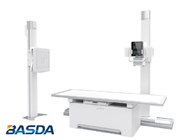
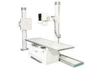
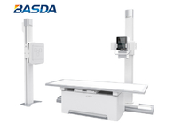
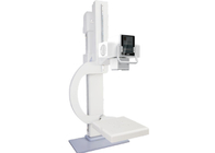
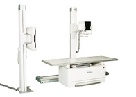
BTR-650B
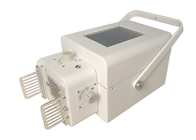
- Digital X-Ray flat panel detector
- Acquisition and processing workstation
- Fully automatic tracking and positioning
- Intelligent control
- Advanced image processing software
- AEC function and DAP function
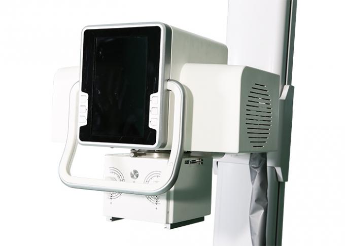
Full feature in-room console
- Selecting the location for photography
- SID
- Tube height
- Detector height
- Angle of column rotation
- mAs value
- mA value
- kV value
- mAs value
- AEC (automatic exposure control)
- Density value
- Selection of large and small focus points
- Exposure Icon
- Detailed information about thermal capacity
Simple design exposure console
- HV on
- HV ready
- HV off
- Exposure ready
- Exposure
Advanced FPD
The full digital detector is a high-performance, full-plate design that overcomes the surface dead zones and pixel shifts that occur when splicing panels together to form line crossings.
No stitching, no artifacts, good stability, long life, good uniformity of image performance, high DQE, fast refresh speed.
Ultra-high-speed image acquisition, from exposure to preview in just seconds, presents the best image at the first time and improves the overall work efficiency.
| |
Full-plate design |
Patchwork plate design |
| Image quality |
1. High fidelity
2. Good image uniformity and stability
|
1. With "dead zone" come from interpolation calculation
2. Poor image uniformity, poor stability
|
| Imaging speed |
Fast |
Slow |
| Life |
>10 years |
<10 years |
Integrated design of wireless plate and battery (some models), no need to remove the battery, reducing the workload of technicians.
The whole board is lighter in weight and stronger in structure, with anti-collision and anti-drop protection design to reduce damage.
AEC
- Air Ionization Chamber is a detector that uses the ionization effect of ionizing radiation to measure the ionizing radiation to achieve automatic exposure control.
- The function. The ionization chamber is divided into three advantageous areas of multiple choice according to the characteristics of the human body.
- The software will preset different advantageous zone combinations on the operation panel of the high voltage generator according to different parts of the film, so there is no need to select them manually.
Rich preset low-dose exposure parameters
The system provides a large number of pre-set exposure parameters, which can be used directly without additional settings, and each group of exposure parameters can be selected as Fat, Medium, or Thin Infant, dramatically improve efficiency while reducing patient radiation and easily obtaining optimal radiation dose -Image quality relationship.
With the easy-to-use collimator, it is easy to obtain high quality exposure images.
① Knob
② Knob
③ Light on/off
④ FOV
⑤ Collimator rotation
⑥ Laser light
Ergonomic software design
- Independent technician accounts: the system can assign a separate account to each operator technician, each of which can store their unique usage habits.
- Including exposure data, sending and printing habits, etc., technicians do not interfere with each other.
- Graphic design APR, objective and clear.
- The software includes modules for patient management, image acquisition, image post-processing, film printing, remote support, etc., which can be used for multiple purposes and effectively save costs for the hospital.
- Image data can be taken and used on traditional media such as CDs, DVDs, portable hard drives, etc., or can be generated in common PC formats such as DICOM protocol supports PACS or RIS, facilitating efficient data management, printing, and other functions., archiving, etc.
Thoughtful detailing
- The system can be configured with a two-way intercom system, which is conducive to real-time communication between technician and patients, and can achieve the recording and playback of different dialects, avoiding the technician repeatedly shouting.
One-stop clinical software package
- The system is equipped with a wealth of image processing functions and management software, including a variety of advanced processing functions to optimize the captured images. the features include the following.
- Patient Management: include simple, advanced or custom data query method; Dicom standard image CD burning for viewing on any standard image workstation; historical image data query and management; disk space detection for automatic cleaning of old examination data; Dicom sending of images, and seamless connection to hospital PACS.
- Software-controlled exposure parameters. Manual/automatic/preset window widths, local widths; image negative/positive, image flip, image rotation, image zoom and Roaming; image information addition; image line, angle, polygon and other measurement tools; organizational equalization, image contrast enhancement, pixels Dose Optimization; Edge Enhancement: Automatically recognizes and analyzes images to enhance image edge sharpness; Image Post-Processing 1:1 Preview: In-doing When image optimized it enables doctors to diagnose more visually.
- Dynamic Range Optimization: include Automatic compression of the original image dynamic curve; high-frequency image enhancement; enhance image contrast, improve the resolution of details, significantly improve the image of bone trabeculae and other images; noise suppression: automatic filtering of cross-frequency signals, significantly reduce image noise and improve the image signal-to-noise ratio.
- Film printing: include film properties, image layout, print method settings; Patient information and display location customization; support for queue management; support for print priority settings.
Unique post-processing optimization techniques
- Unique post-processing optimization technique spatially fills the original image captured by the detector, improving data integrity makes the edges of the tissues sharper, the tissue hierarchy better, the structure is clearer and the image uniformity is more consistent.
- Taking the region of complex bony structures as an example, the pre-optimization (left) and post-optimization (right) results are shown below.
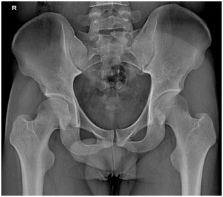
Automatic background recognition removal technology
- The software automatically identifies and removes artifacts and grids outside the anatomical structures of various parts of the human body, maximizing effective tissue contrast and resolution.
- The images quality are shown below.
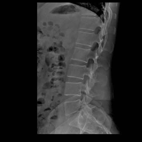
Optimized full spinal stitching function
- Self-developed algorithm and technology, can easily complete the seamless stitching of the whole spine imaging, overcoming the inconsistent gray and dark contrast of the traditional stitching image. The image quality is shown below.
Client Training
- Equipment function introduction;
- Operation manual introduction;
- Device maintenance;
- Efficient use ways of the device.
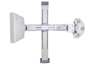
BTR-650B Configuration
| No. |
Part |
Specification |
| |
Power input |
220V, 5kW |
| 1 |
High voltage generator |
Power output: 50kW |
| Max. kV value: 150kV |
| Max. mA value: 630mA |
| Min. exposure time: 1ms |
| Min. mAs: 0.4mAs |
| With APR function |
| HV console at workstation |
| With AEC link function |
| Inverter frequency: 100kHz |
| 2 |
Console |
CPU :3.2GHz
OS:win 7
RAM:8GB
HDD:1000GB
DVD recorder: 4.5GB
Ethernet: 100BaseT/1000BaseT
Comply DICOM3.0 including print, storage, CD/DVD, transfer / receive and worklist
LCD size:≥19"
Mouse and keyboard
Patient register module
Exposure parameter module
3D APR function
Image view and process module
Print module
|
| 3 |
Tube |
Brand: Toshiba |
| Anode rotation speed: 9000rpm |
| Max peak X-ray tube voltage: 150KV |
| Anode heat storage capacity: 300kHU |
| Focal spot: 0.6mm/1.2mm |
| 4 |
Gantry |
Type: dual column |
| With auto tracking function |
| Tube console parameter: exposure, SID, tube angle, focal spot, AEC |
| Tube console size: 10.1” |
| Detector support longitudinal movement range:≥1800mm |
| Detector support vertical movement range:≥1000mm |
| Tube support rotation range ≥360° |
| 5 |
Collimator |
Manual control
Coherent filter: 1.0-1.3mmAL
Adjustable filter: 0.5/1.0mmAL
Min. exposure field: 10mm×10mm
Max. exposure field: 430mm×430mm;
|
| 6 |
Detector |
Material: ASi-CsI |
| Mobile type |
| Min. pixel size: 140um |
| A/D: 16bit |
| Space resolution: 3.6lp/mm |
| Preview time: 3s |
| Detector size: 17” * 17” |
| Matrix: 3000 * 3000 |
| 9 |
Cable |
>9m |
| 10 |
Patient table |
Floating table with magnetic lock
Table longitudinal float range:900mm
Table horizontal float range:250mm
Table size: 2300mm×810mm;
Table height: 680mm
Table support capacity≥200kg
Grid density: 40L/cm
Grid size: 450 * 450mm
With 3 field AEC
Detector movement range: 1000mm
|
 Your message must be between 20-3,000 characters!