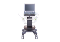
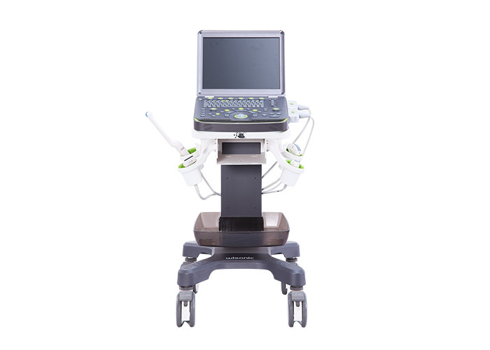
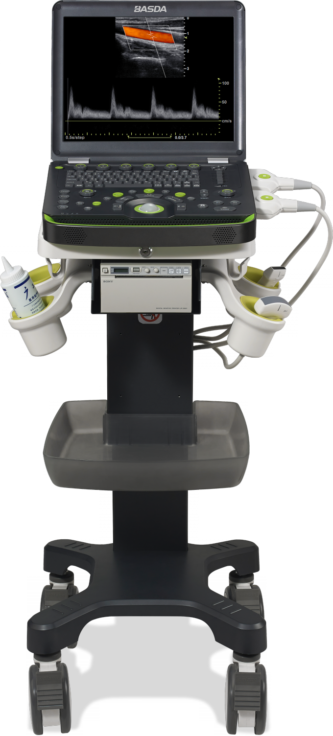
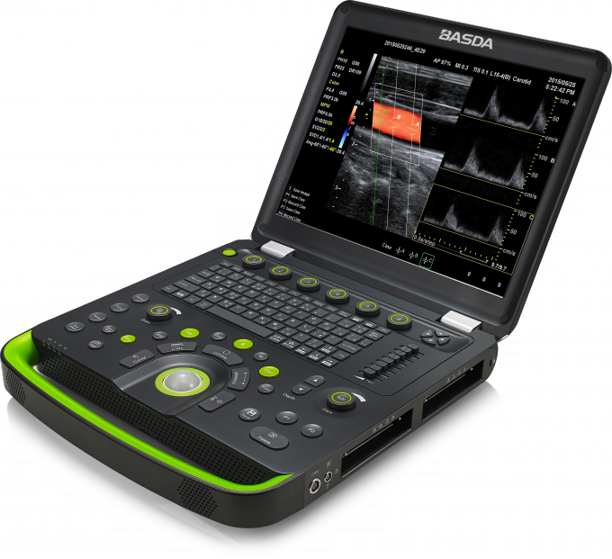
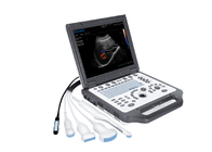
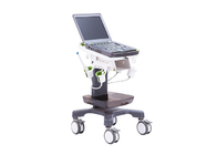
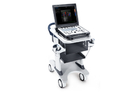
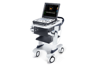
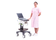
BTH-50S Portable Color Doppler Ultrasound doppler 3D 2 probe sockets large volume battary easy track ball
1 Application
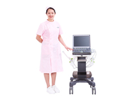
It is mainly applied for ultrasonic diagnosis and scientific research of abdomen, obstetrics and gynecology, reproduction, blood vessels, heart, superficial tissue, breast, neonates, surgery, nerves, etc.
2 Specification:
2.1 Include:
High resolution B imaging
2.2 Measurement and analysis:
2.2.1 Preset parameters: for different examined organs, the optimal image examination parameters are preset to reduce the adjustment during operation; users are permitted to define and save preset parameters
2.2.2 General measurement
2.2.3 Obstetrics measurement(evaluation of fetal weight and gestational age, displaying custom growth curve, measurement of fetal cardiogram)
2.2.4 Gynecology measurement
2.2.5 Vessel measurement
2.2.6 Cardiac measurement
2.2.7 Abdomen measurement
2.2.8 User defined measurement
2.2.9 Doppler blood flow measurement and analysis
2.2.10 Small organs and superficial tissue measurement
2.2.11 Auto doppler spectrum tracking and analysis
2.2.12 Auto measurement of intima-media thickness
2.2.13 Assigned measurement function: items can be defined and results annotated
2.3 Patient data management:
2.3.1 Patient data include: personal information, image, cine, report, etc
2.3.2 Patient data establishment, storage, playback, modification, retrieval and printing
2.3.3 Built-in integrate ultrasound workstation
2.3.4 Image storage format: PNG, JPEG, BMP
2.3.5 Cine storage format: AVI
3 Technical parameters:
3.1 System general configure:
3.1.1 Full digital uwb beamformer: digital channels ≥ 28672
3.1.2 A/D sampling ≥ 14 BIT
3.1.3 15 inch high resolution LED color monitor
3.1.4 Net weight built-in battery: ≤ 5 kg
3.1.5 Operation panel:physical keyboard and mechanical trackball
3.1.6 *Dual transducer connectors, non-extended connector
3.1.7 Battery life on real-time scanning: ≥ 70 min
3.1.8 SSD≥250G
3.1.9 Interfaces: network, HDMI, S-video, VGA,USB
3.1.10 Support U disk or mobile HDD storage
3.1.11 Support black white printer and color printer
3.1.12 Optional special trolley with storage box
3.2 Transducer specification:
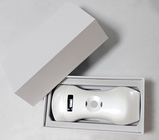
3.2.1 Support transducer type: convex array, linear array, phased array, and endocavity
3.2.2 The maximum number of array elements: ≥ 192
3.2.3 Transducer supports biopsy guide
3.2.4 Frequency range:
- Convex array transducer frequency: 2.5-5.5 MHz
- Phased array transducer frequency: 2.5-4.5 MHz
- Linear array transducer frequency: 5.0-12.0 MHz
- Endocavity transducer frequency: 5.0-8.0MHz
3.2.5 Imaging modes:
- Convex array transducer: B/M/CDI/PWD
- Linear array transducer: B/M/CDI/PWD
- Endocavity transducer: B/M/CDI/PWD
- Phased array transducer: B/M/CDI/PWD/CWD
3.3 B imaging parameters:
3.3.1 Display mode: B, B/B,4B, M,B/M
3.3.2 Gain adjustment: TGC ≥8; LGC ≥2
3.3.3 Transmit beam focusing: ≥ 4
3.3.4 Gray scale: ≥ 256
3.3.5 Scanning depth: ≥ 29cm
3.3.6 Visual and adjustable dynamic range: 30 ~ 180dB
3.3.7 Cineloop: B images playback ≥16000 frames
3.3.8 B scanning rate:
- phased array transducer: when depth is 18 cm, the full-field scanning
- frame rate ≥70 f/s
- convex array transducer: when depth is 18 cm, the full-field scanning
- frame rate ≥79 f/s
3.4 Color doppler imaging parameters:
3.4.1 Type: CDI, PDI, Dir PDI
3.4.2 Display modes: B/CDI, B/PDI, B/Dir PDI
3.4.3 B and color dual real-time contrast
3.4.4 Zero movement: ≥8 levels adjustable
3.4.5 Steer angle: linear array transducer image steer range: -30°~ +30°
3.4.6 Color scanning rate:
- phased array transducer: when depth is 18 cm, the full-field scanning frame rate ≥ 16 f/s
- convex array transducer: when depth is 18 cm, the full-field scanning frame rate ≥ 8 f/s
3.5 Exclusive Imaging Patents:
3.5.1 ePure
- Intelligent speckle noise suppression technology can intelligently identify different tissue information in different spatial dimensions point-to-point, suppress the edge information display of speckle noise, and make the image more exquisite.
3.5.2 eSCI
- Electronic space composite imaging technology can update the echo signals received from different angles in real time through multi-angle deflection scanning in space and continuously update fusion imaging. On the premise of ensuring the time resolution.
3.5.3 eFCI
- The function can automatically identify and measure the thickness of endovascular media. At the same time, the measurement report of inner and middle membrane thickness is provided to users with more accurate and more efficient assessment information of vascular diseases.
3.5.4 eView
- Ultra-wideband Imaging with expansion of the special tissue anatomical structure through visualization to provide more and more complete tissue relationship information.
3.5.5 eSpeed
- The function could help process clear images automatically depending on different exam modes which could enhance the efficiency for users.
 Your message must be between 20-3,000 characters!