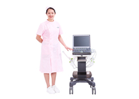
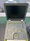
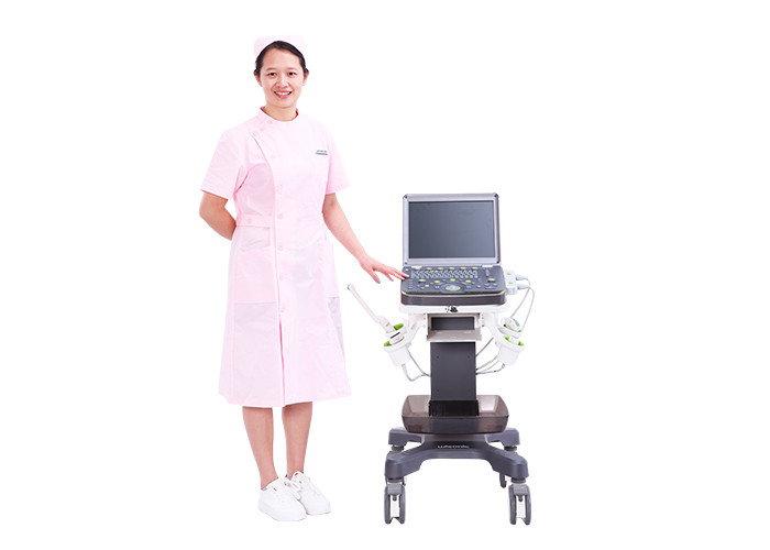
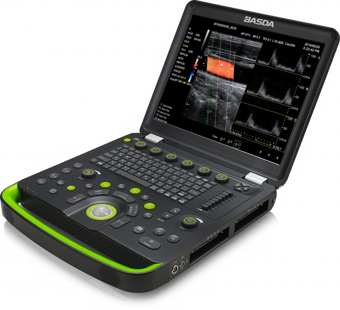
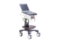
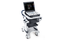
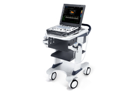
BTH-50S Portable Color Doppler Ultrasound doppler spectrum 2 probe sockets large volume battary
1 Application
It is mainly applied for ultrasonic diagnosis and scientific research of abdomen, obstetrics and gynecology, reproduction, blood vessels, heart, superficial tissue, breast, neonates, surgery, nerves, etc.
2 Specification:
2.1 Include:
2.1.1 High resolution B imaging
2.1.2 Color doppler imaging (CDI )
2.1.3 Pulse wave doppler (PWD)
2.1.4 Continuous wave doppler (CWD)
2.1.5 Power doppler imaging (PDI)
2.1.6 Direction Power doppler imaging (DirPDI)
2.1.7 M mode imaging
2.1.8 Multi angle M imaging
2.1.9 Tissue harmonic imaging
2.1.10 Real-time spacial composite imaging, both used for transmit and receive, ≥7 lines
2.1.11 Adaptive imaging enhancement, clear Speckle noise, improve resolution of tissue boundary contrast.
2.1.12 Frequency composite imaging, the transmitting frequency is adjusted adaptively and composited according to depth.
2.1.13 *Three PWD sampling gates and three PWD imaging appear simultaneously within one heart cycle
2.1.14 *Auto doppler blood vessel tracing: Auto locates the blood vessels, then adjusts the color ROI box position, steer angle, gate size, PWD steer angle and correction angle
2.1.15 Smart one key imaging optimization: optimize B, CDI, PWD imaging
2.1.16 Zoom in full screen
2.1.17 Real-time zoom
2.2 Measurement and analysis:
2.2.1 Preset parameters: for different examined organs, the optimal image examination parameters are preset to reduce the adjustment during operation; users are permitted to define and save preset parameters
2.2.2 General measurement
2.2.3 Obstetrics measurement(evaluation of fetal weight and gestational age, displaying custom growth curve, measurement of fetal cardiogram)
2.2.4 Gynecology measurement
2.2.5 Vessel measurement
2.2.6 Cardiac measurement
2.2.7 Abdomen measurement
2.2.8 User defined measurement
2.2.9 Doppler blood flow measurement and analysis
2.2.10 Small organs and superficial tissue measurement
2.2.11 Auto doppler spectrum tracking and analysis
2.2.12 Auto measurement of intima-media thickness
2.2.13 Assigned measurement function: items can be defined and results annotated
2.3 Patient data management:
2.3.1 Patient data include: personal information, image, cine, report, etc
2.3.2 Patient data establishment, storage, playback, modification, retrieval and printing
2.3.3 Built-in integrate ultrasound workstation
2.3.4 Image storage format: PNG, JPEG, BMP
2.3.5 Cine storage format: AVI
3 Technical parameters:
3.1 System general configure:
3.1.1 Full digital uwb beamformer: digital channels ≥ 28672
3.1.2 A/D sampling ≥ 14 BIT
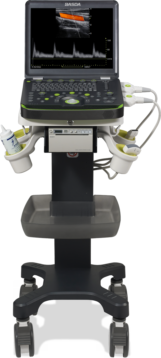
3.1.3 15 inch high resolution LED color monitor
3.1.4 Net weight built-in battery: ≤ 5 kg
3.1.5 Operation panel:physical keyboard and mechanical trackball
3.1.6 *Dual transducer connectors, non-extended connector
3.1.7 Battery life on real-time scanning: ≥ 70 min
3.1.8 SSD≥250G
3.1.9 Interfaces: network, HDMI, S-video, VGA,USB
3.1.10 Support U disk or mobile HDD storage
3.1.11 Support black white printer and color printer
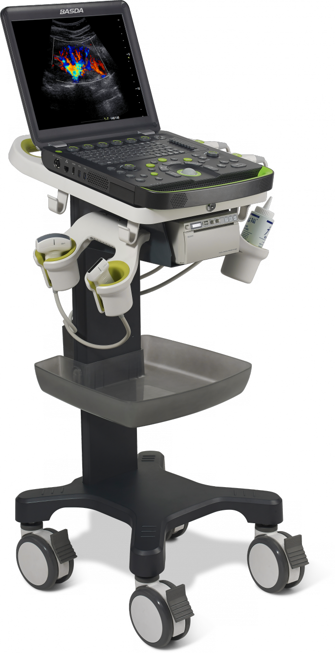
3.1.12 Optional special trolley with storage box
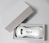
3.2 Transducer specification:
3.2.1 Support transducer type: convex array, linear array, phased array, and endocavity
3.2.2 The maximum number of array elements: ≥ 192
3.2.3 Transducer supports biopsy guide
3.2.4 Frequency range:
- Convex array transducer frequency: 2.5-5.5 MHz
- Phased array transducer frequency: 2.5-4.5 MHz
- Linear array transducer frequency: 5.0-12.0 MHz
- Endocavity transducer frequency: 5.0-8.0MHz
3.2.5 Imaging modes:
- Convex array transducer: B/M/CDI/PWD
- Linear array transducer: B/M/CDI/PWD
- Endocavity transducer: B/M/CDI/PWD
- Phased array transducer: B/M/CDI/PWD/CWD
3.3 B imaging parameters:
3.3.1 Display mode: B, B/B,4B, M,B/M
3.3.2 Gain adjustment: TGC ≥8; LGC ≥2
3.3.3 Transmit beam focusing: ≥ 4
3.3.4 Gray scale: ≥ 256
3.3.5 Scanning depth: ≥ 29cm
3.3.6 Visual and adjustable dynamic range: 30 ~ 180dB
3.3.7 Cineloop: B images playback ≥16000 frames
3.3.8 B scanning rate:
- phased array transducer: when depth is 18 cm, the full-field scanning
- frame rate ≥70 f/s
- convex array transducer: when depth is 18 cm, the full-field scanning
- frame rate ≥79 f/s
3.4 Color doppler imaging parameters:
3.4.1 Type: CDI, PDI, Dir PDI
3.4.2 Display modes: B/CDI, B/PDI, B/Dir PDI
3.4.3 B and color dual real-time contrast
3.4.4 Zero movement: ≥8 levels adjustable
3.4.5 Steer angle: linear array transducer image steer range: -30°~ +30°
3.4.6 Color scanning rate:
- phased array transducer: when depth is 18 cm, the full-field scanning frame rate ≥ 16 f/s
- convex array transducer: when depth is 18 cm, the full-field scanning frame rate ≥ 8 f/s
3.5 Spectral doppler imaging parameters:
3.5.1 Type:PWD,CWD,HPRF
3.5.2 Display modes: B/PWD, B/CWD, B/HPRF, B/CDI/PWD, B/CDI/CWD, duplex/triplex
3.5.3 The maximum blood flow velocity : ≥8.5 m/s by PWD ; ≥35 m/s by CWD
3.5.4 The lowest blood flow velocity:≤3 mm/s (non-noise signal)
3.5.5 Cineloop:≥ 400s,cineloop of doppler mode and M mode can be measured and calculated
3.5.6 Zero movement: ≥8 levels
3.5.7 Sampling width and position range: 1- 30 mm grading
4 Fast Description
- Highly Mobile, Slim and Smart Design
- Quick Boot Up Time
- Built-In Battery Supports Longer Duration
- Smart Design with 2 standard probe ports
- High Performance Transducer: Abdominal, Cardiac, Intracavitary, Small Parts
5 Advanced Technology
- Holographic Ultrasound Platform
- Triple Sampling Gates of PW (Pulse Wave) Doppler
- Speckle Noise Suppression Technology
- Space and Frequency Composite Imaging
- One-Click Automatic Optimization
- Automatic Tracking and Measurement
- Ultra-rapid Intelligent Search
6Client Training
1) Equipment function introduction;
2) Operation manual introduction;
3) Device maintenance;
4) Efficient use ways of the device.
 Your message must be between 20-3,000 characters!