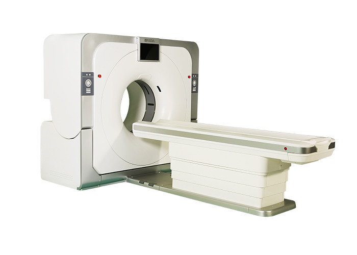
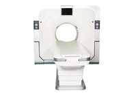
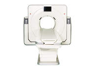
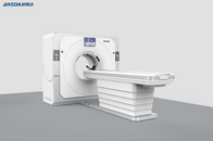
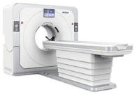
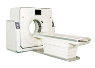
BCT - 32
CT scan,Computed Tomography system,32 slices detector,76cm,5.3MHU
• BCT - 32 is a new 32 slice spiral CT scanner that BASDA company introduced in 2016, the system includes the industry's top technology, it can produce high-definition images, it has rich software function, meets the demand of the hospital for large daily patient flow, it has the major characteristics include:
• The image chain of BCT-32 adopts mature technologies, the integrity of the system is balanced and strong.
• The system config the industry's leading dynamic real-time zooming technology and real 3D cone parallel reconstruction technology, so that the system can get the aperture expanded and at the same time get high definition image and leading high resolution image quality.
BCT-32 includes below high-lighted technologies and features:
Mature and stable large heat capacity tube
• This tube can meet the demand of large patient flow and complex scanning, in the meantime, guarantee the system operating costs under control.
Dynamic reconstruction convolution technology based on FOV
• Dynamic reconstruction convolution technology based on FOV can real-time match the algorithm along with the different field of view to ensure different tissues have an optimal effect in the images, this technology is similar as the digital camera, ensures that the users can dynamically capture the best photos.
- The upper left and right image are identical and replace them with a better patient body image generated by our CT at FOV of 50cm, 45cm or 40cm, whichever is appropriate.
- The lower left image was just cut off the upper left image into half in size and enlarge by factor 2 to get the same size but blurred image.
- Replace the blank blue block with the image reconstructed at half FOV of the upper right image, using the image matrix dimension 512x512 identical to that of the upper right image.
- As such, the point of dynamic reconstruction kernel can be illustrated.
Intelligent mA tracking technology
• On the premise of the quality of the image guaranteed, the system can automatically adjusts the output tube current in Z axis direction and X axis direction. Intelligent mA tracking technology can reduce about 40% radiation dose.
Parallel 3D cone beam reconstruction technology
• Integrated parallel computing reconstruction based on true 3D cone beam, the final image is closer to the true tissue, the image is clearer.
iDream tensor iteration low-dose technology
76 cm aperture design
• Place the patients at ease and comfortable.
| No. |
Item |
Specification |
| 1 |
Gantry |
|
| 1.1 |
Patient Opening |
76cm |
| 1.2 |
Gantry Driven Type |
Motor-driven |
| 1.3 |
Tilt Capability |
±30° |
| 1.4 |
Gantry Remote-Control |
Yes |
| 1.5 |
Detector Type |
GOS high-definition solid detector |
| 1.6 |
Distance of Focus-Isocenter |
570mm |
| 1.7 |
Numbers of Detector Rows |
32 |
| 1.8 |
Width of Z-Axle Detector |
20mm |
| 1.9 |
Detector Columns of Channels per Row |
864 |
| 1.10 |
Numbers of Detector Columns |
27648 |
| 1.11 |
Slice Number |
32 |
| 1.12 |
DAS Column Number |
55296 |
| 1.13 |
DAS Acquisition Modes |
32×0.625mm
16×1.25mm
8×2.5mm
4×5.0mm
2×10mm |
| 1.14 |
Data-Transfer Type |
RF,optical fiber communication |
| 1.15 |
3D Laser Orientation |
Provided |
| 1.16 |
Automatic Exposure Control |
Provided |
| 1.17 |
Auto-Voice Function |
Yes |
| 2 |
Scan Parameter |
|
| 2.1 |
Shortest 360 Degree Rotation Time |
0.5s |
| 2.2 |
Allowed Rotation Times |
0.5s,0.6s,0.7s
0.8s,1.0s,2.0s
|
| 2.3 |
Slice Numbers of Rotation |
32 |
| 2.4 |
Minimum Scan Slice Thickness |
0.625mm |
| 2.5 |
Minimum Reconstruction Thickness |
0.625mm |
| 2.6 |
Maximum Scan Slice Thickness |
10mm |
| 2.7 |
Reconstruction Slice Thickness |
0.625mm |
| 2.8 |
Speed of Image Reconstruction |
40 frames/s |
| 2.9 |
Scan FOV |
50cm |
| 2.10 |
Max. Image Reconstruction Matrix |
1024×1024 |
| 2.11 |
Max. Image Display Matrix |
1024×1024 |
| 2.12 |
Maximum Continuous Scan Duration |
100s |
| 2.13 |
Maximum Continuous Scan Length |
1950mm |
| 2.14 |
Range of Pitch |
0.13-1.5 |
| 2.15 |
Scan Mode |
Scout Scan |
| Axial Scan |
| Helical Scan |
| Cine Scan |
| 3 |
HV generator and Tube |
|
| 3.1 |
Maximum Output Power of Generator |
50kW |
| 3.2 |
Tube kV Selections |
80kV,100kV
120kV,140kV
|
| 3.3 |
Tube mA Range |
10~420mA |
| 3.4 |
Tube Heat Capacity |
5.3MHu |
| 3.5 |
Heat Dissipation Rate |
815 kHU/min |
| 3.6 |
Type of Cooling |
Oil cooling + Air cooling |
| 3.7 |
Tube Focus |
1.1mm×1.1mm
0.5mm×1.0mm
|
| 3.8 |
Flying Focal Spot Technology |
Provided |
| 3.9 |
mA Modulation Technology |
Provided |
| 4 |
Patient Table |
|
| 4.1 |
Can Position Outside of Scan Room |
Yes |
| 4.2 |
Laser Position System Accuracy |
±1mm |
| 4.3 |
Maximum Horizontal Movable Range |
1950mm |
| 4.4 |
Table Horizontal Scan Range |
1850mm |
| 4.5 |
Table accuracy |
0.25mm |
| 4.6 |
Table Vertical Movable Range |
425~990mm |
| 4.7 |
Maximum Speed of Vertical Movement |
16.5mm/s |
| 4.8 |
Maximum Speed of Horizontal Movement |
5 mm/s ~ 160 mm/s ±3% |
| 4.9 |
Maximum Patient Weight |
250kg |
| 4.10 |
Foot Pedal of Table |
Optional |
| 5 |
Image Quality |
|
| 5.1 |
High Contrast Resolution |
≥20lp/cm
(0% MTF)
|
| 5.2 |
Low Contrast Resolution |
3.0mm@0.30%
(31mGy)
|
| 5.3 |
Isotropic Imaging Resolution |
0.3mm |
| 5.4 |
Range of CT Value |
-1000~+1000 |
| 5.5 |
Image Noise |
≤70dB |
| 6 |
Computer |
|
| 6.1 |
CPU |
4coreX3.5Hz |
| 6.2 |
Memory |
32GB |
| 6.3 |
Storage of Hard-Disk |
5TB |
| 6.4 |
Monitor |
24’’ LCD Monitor |
| 6.5 |
Resolution of Monitor |
1920 x 1200 |
| 6.6 |
Image-Data External Storage Type |
CD/DVD/USB |
| 6.7 |
DICOM 3.0 |
Provided |
| 6.8 |
Worklist |
Provided |
| 6.9 |
3D Post-Processing |
MPR,CPR,SSD,MIP,VR,CTA |
| 6.10 |
Contrast Agent Automatically Tracking and Trigger Technology |
Provided |
| 6.11 |
X-ray intelligent control technology |
Provided |
| 6.12 |
Maximum CTA Scan Range |
1850mm |
| 6.13 |
Beam Artifacts Correction Technology |
Provided |
| 6.14 |
Posterior Fossa Image Optimization Technology |
Provided |
| 6.15 |
Metal Artifacts Algorithm |
Provided |
| 6.16 |
Scalpel Simulate Technology |
Provided |
| 6.17 |
Child Mode |
Provided |
 Your message must be between 20-3,000 characters!