Home ProductsBasda MRI

Water Cooling Method Basda MRI Machine 1.5 T Superconducting 60cm Width Bstar-150

Water Cooling Method Basda MRI Machine 1.5 T Superconducting 60cm Width Bstar-150

Water Cooling Method Basda MRI Machine 1.5 T Superconducting 60cm Width Bstar-150
Product Details:
Place of Origin: China
Brand Name: BASDA
Certification: CE, ISO13485
Model Number: Bstar-150
Payment & Shipping Terms:
Minimum Order Quantity: 1 set
Price: Negotiable
Packaging Details: 1 unit/wooden box
Delivery Time: 30-45 Days
Payment Terms: Credit card, Paypal, T/T, Western Union, MoneyGram
Supply Ability: 200 Machines per Year
Contact Now
Detailed Product Description
Cooling Technology: Water Cooling Method Machine Material: Liquid Helium Magnet
Min. 3D Thickness: 0.05 Mm Bore Diameter Size: 60 Cm Width
Patient Table Length: Around 2.4 Meters Load Of Patient Table: Less Than 200kg
High Light:

Superconducting Basda MRI Machine

,

1.5 T Basda MRI Machine

,

Water Cooling 1.5 T Mri Machine

 
What to Expect from 1.5 Tesla Superconducting MRI with 800L Liquid Helium
  • BASDA superconductive MRI Bstar-150 is the one that break the technology monopoly by foreign companies, a new generation of superconductive MRI that based on rich research and development and clinical experience, is applicable to all parts of the body, fully meet the central nervous system, abdominal, pelvic cavity and etc.
  • Golden-ratio design magnet. A long magnet will make the patient feel depressed during the examination while a short magnet will greatly decrease the homogeneity. The "golden-ratio" design magnet is the elegant balance between both.
  • The smart control screen mounted on the gantry, helps the operator to confirm the table position and patient status in real time, increasing the daily patient flow.
  • With high accuracy diagnosis automatic bed design and intelligent speed control, it has greatly improved patient comfort as well as patient flow.


Bstar-150 Contains a Variety of Coils
 
The Receiver coil, together with parallel acquisition technology, improve the signal-to-noise ratio and image quality. The customer can choose according to the clinical needs (specific configuration of the coil number and types will be subject to final contract).
  • AIT coil set
  • Head coil
  • Neck coil
  • Body coil
  • Knee coil
  • Shoulder coil
  • Ankle coil
  • Wrist coil
  • Breast coil
 
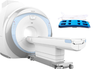
1.5T Superconducting MRI  Software
 
RF sub system
 
The RF sub system is the source of MRI proton excitation energy, its efficiency directly affects the MRI relax signal. RF receiver receive MRI relax signal, a good receiving coil can improve the SNR of image.
 
The RF sub system is similar as highway lane, the more lanes, the faster flow of vehicles. Vice versa, the more the RF system channels, the faster parallel acquisition, thus the system can process the greater the amount of data in the same time, reduce the imaging time. Bstar-150 suing 8 / 16 channel radio frequency receiving, combined with the latest parallel sampling technology, using the DDC digital direct sampling, can short the scan time and get excellent image quality.
 

Bstar-150 provides a versatile suite of imaging capabilities for body imaging to meet the growing demands of abdominal, pelvic, and breast exams. Including MRU/MRCP/MRM imaging, and other advanced imaging sequences. Rich post-processing software can optimize images in case necessary to help in complex diseases diagnosis, excellent image quality and abundant imaging techniques.

 
Gradient sub system
 
Gradient sub system directly determines the spatial encoding of MRI system, it produces the signal information, at the same time it can perform GR series sequence scanning, it is the foundation of MRI system’s spatial resolution ability, gradient sub system also directly affect the scanning image thin slice scanning.
 
The linearity of gradient system of MRI system determine the accuracy of spatial location information, ensuring that in any acquisition, the image distortion is limited.
 
Gradient switching rate is an important factor of MRI scanning speed. Bstar-150 using the self-shielding gradient coil and high duty cycle gradient amplifier system, together with eddy elimination technology and design, can avoid the eddy current between the metal shielding layers, raise up the spatial encoding ability of the magnetic resonance signal and make the scan more precise, ensure high resolution image quality.
 
During superconducting MRI scans, the noise is much bigger than permanent MRI systems, and staying in a closed environment, the patient is easy to fall into tension. Bstar-150’s gradient sub system adopts special hardware noise reduction design technology, the gradient noise is reduced to 2/3, provide patients with friendly scanning environment.
 
The gradient system is equipped with automatic detection function, if the temperature is too high, the system can intelligently stop the scanning, to protection of hardware from being damaged.
The system use water chiller to produce a stable running condition.


Workflow is critical to the efficient operation of an MRI. It impacts patient satisfaction, image quality and cost effectiveness. Bstar-150 provides full preset of clinical diagnostic scan protocol, based on rich clinical experience, corresponding arrangement and optimization have been made per related clinical application features for the scan procedure and sequence parameters of the preset scan method. With unmatched clinical capability, Bstar-150 provides an unprecedented ability to acquire complete exams with a motion compensated scan technique.


High resolution isotropic diffusion imaging, based on powerful gradient system and amplifier system, Bstar-150 realizes faster and more reliable DWI images with high resolution, anatomic structure of head is clear, diffusion image features is obvious, very convenient for lesion location lock and lesion nature diagnosis.HD susceptibility-weighted imaging, imaging based on the differences of magnetic susceptibility in different tissues, is a contrast enhancement technique that reflects magnetic properties of tissue. It is extremely sensitive to bleeding or deoxidation part in blood, can provide accurate information on bleeding, arteriovenous malformation, iron deposit to realize faster and more accurate diagnosis, even the very small lesion can be quickly diagnosed too.


Fast high-resolution abdomen diffusion imaging, under the support of a brand-new parallel acquisition technique, realizes abdomen diffusion imaging with high spatial resolution, high time resolution and high contrast resolution, providing an excellent tool of abdomen cancer diagnosis.


Extremely HD 3D TOF blood vessel imaging, optimized K space acquisition and filling method, suppression uniform of obtained images background, no artifact, clear blood vessel trend, can display tertiary vascular or above.


scDWI (optional)

In DWI, acquisition of low-b-value has better tissue SNR, but is not sensitive enough to water proton;


Acquisition of high-b-value has better DW sensitivity of water proton, but reduces the tissue SNR as well as the lesion location function;


By using special computation method, scDWI only needs one single DWI sequence to complete the calculation of multi-b-value within certain range, min. 100s/mm².


Meanwhile, comparing to direct acquisition of DWI images, scDWI technique greatly improved the SNR of high-b-value DW image, avoid distortion of high-b-value images, very good for lesion diagnosis.


Bstar-150 Specification


No. Item Specification
1 Magnet System  
1.1 Magnet Type Superconducting
1.2 Field Strength 1.5T
1.3 Shielding Method Active
1.4 Shimming Method and Type Active + Passive + Dynamic
1.5 Magnet stability ≤0.1ppm/h
1.6 Homogeneity (DSV, VRMS)  
1.6.1 45cm ≤0.832ppm
1.6.2 40cm ≤0.323ppm
1.6.3 30cm ≤0.107ppm
1.6.4 20cm ≤0.041ppm
1.6.5 10cm ≤0.01ppm
1.7 Length of magnet(exclude cover) 157cm
1.8 Inner diameter of magnet 600mm
1.9 5 Gauss fringe field(X,Y,Z axis) ≤2.5m x 2.5m x 4.0m
1.10 Total capacity of liquid helium (100% liquid helium full filled) 800L
1.11 Filling liquid helium period ≥4 Years
1.12 Liquid helium “zero”consumption technology Yes
2 Gradient System  
2.1 Maximum gradient field (single axis, invalid) 41mT/m
2.2 Maximum gradient slew rate(single axis, invalid) 187mT/m/ms
2.3 Minimum gradient rise time 0.22ms
2.4 Maximum gradient field and slew rate reached at the same time Yes
2.5 Gradient cooling system Water cooling
2.6 Full digital real-time transmit and receiving gradient control system Yes
3 RF System  
3.1 Max RF amplifier power 18kW
3.2 Center frequency 63.87MHz
3.3 Real-time digital RF energy monitoring Yes
3.4 Real-time digital RF short-term accumulation monitoring Yes
3.5 Real-time digital RF long-term accumulation monitoring Yes
3.6 Parallel working RF receiver channels 8/16
3.7 Parallel working RF A/D converters 8/16
3.8 Each coil unit has corresponding pre-amplifier Yes
3.9 Parallel acquisition technology platform Yes
3.10 Maximum bandwidth of each parallel acquisition receive channels ≥1.0MHz
3.11 Transmission bandwidth 550kHz
3.12 Maximum receiver signal resolution ≥16bit
3.13 Fully digital RF system Yes
3.14 RF receiving amplifier noise level ≤0.45dB
3.15 Multi-channel phased array receiver coil  
3.15.1 Head coil Available
3.15.2 Neck coil Available
3.15.3 Body coil Available
3.15.4 Knee coil Available
3.15.5 Shoulder coil Available
3.15.6 Ankle coil Available
3.15.7 Wrist coil Available
3.15.8 Breast coil Available
  Remark: The coil configuration is subject to the final contract  
4 Scan environment  
4.1 Patient table driven mode Motor-driven
4.2 Position accuracy ≤0.5mm
4.3 Table length 2400mm
4.4 Horizontal movement range 2055mm
4.5 Horizontal motion maximum speed ≥200mm/s
4.6 Patient table lowest height 675mm
4.7 Maximum patient weight 200kg
4.8 Feet-first entry mode Yes
4.9 Table can be controlled by machine cover in case of emergency Yes
4.10 Table control system on both side of rack Yes
4.11 Lighting, ventilation, call system Be able to adjust
5 Computer system  
5.1 MRI software BASDA
5.2 System software WINDOWS 7
5.3 CPU ≥3.1GHz
5.4 Main memory ≥4GB
5.5 Hard disk ≥500GB
5.6 Color LCD monitor 24”
5.7 Monitor resolution 1920×1200
5.8 External storage DVD/USB
5.9 DICOM3.0 Yes
6 Scanning parameter  
6.1 Spin-echo sequence Yes
6.1.1 SE 2D/3D Yes
6.1.2 FSE 2D/3D Yes
6.1.3 FSE sharing Yes
6.1.4 Single shot FSE Yes
6.1.5 Spin echo fat-suppression imaging Yes
6.1.6 Spin echo frequency fat suppression imaging Yes
6.1.7 Spin echo water suppression imaging Yes
6.2 GRE 2D/3D Yes
6.3 DWI Yes
6.3.1 Max. b value 10000
6.4 IR sequence Yes
6.4.1 IR Yes
6.4.2 FIR(water / fat suppression) Yes
6.4.3 FLAIR Yes
6.4.4 STIR Yes
6.4.5 Water-fat Separation Yes
6.5.1 Pre-saturation Yes
6.6 Gating Yes
6.7 Accelerated sequence Yes
6.8 Anti-movement scanning technology Yes
6.9 Min. 2D thickness 0.1mm
6.10 Min. 3D thickness 0.05mm
6.11 Max. FOV 50cm
6.12 Min. FOV 1cm
7 Advanced imaging technology  
7.1 Body Imaging  Yes
7.1.1 Ultra-fast imaging technology Yes
7.1.2 Phase / de-phase imaging technology Yes
7.1.3 MR cholangiopan-creatography (MRCP) Yes
7.1.4 MR urography (MRU) Yes
7.1.5 MR Myelography (MRM) Yes
7.2 Neuro imaging Yes
7.3 Diffusion weighted imaging set Yes
7.3.1 Isotropic acquisition Yes
7.3.2 ADC measurement Yes
7.4 MR angiography(MRA) Yes
7.4.1 2D/3D TOF technology Yes
7.4.2 Continuous multi-layer 3D TOF technology Yes
7.4.3 Contrast enhanced MRA Yes
7.4.4 Magnetization transfer (MTC) Yes
7.4.5 Maximum intensity projection Yes
7.5 Susceptibility weighted imaging (SWI) Yes
7.6 Parallel acquisition technology Yes
7.6.1 Algorithm based on image Yes
7.6.2 Algorithm based on K-space Yes
7.6.3 Max. parallel acquisition acceleration factor 4
7.6.4 Automatic calibration technology Yes
7.6.5 Applied direction of parallel acquisition factor X,Y,Z
7.7 Artifact correction technology Yes
7.7.1 Fluid compensation Yes
7.7.2 Respiratory compensation Yes
7.7.3 Head motion artifact correction Yes
7.7.4 Elimination of magnetic sensitive artifact Yes
7.7.5 Eddy current adaptive correction Yes
7.7.6 Gradient linearity correction Yes
7.7.7 Multi-echo phase correction Yes
7.8 Rapid automatic correction technology Yes
7.9 Automatic frequency tracking technology Yes
7.10 Automatic coil recognition technology Yes
7.11 Automatic phase correction technology Yes
7.12 Echo navigation technology Yes
7.13 RF balance drive technology Yes
7.14 RF de-phase technology Yes
7.15 Gradient de-phase technology Yes
7.16 Section scanning technology Yes


Contact Details
Shenzhen Basda Medical Apparatus Co., Ltd

Contact Person: Peng

Tel: +86-13632708845

Send your inquiry directly to us (0 / 3000)

Contact

Address: Floor 19, Block 1A, Basda Building, No. 28 Nantong Road, Longgang District, Shenzhen, Guangdong Province, P.R.C.

Factory Address:Block 1B, Basda Building, No. 28 Nantong Road, Longgang District, Shenzhen, Guangdong Province, P.R.C.