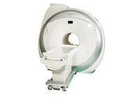
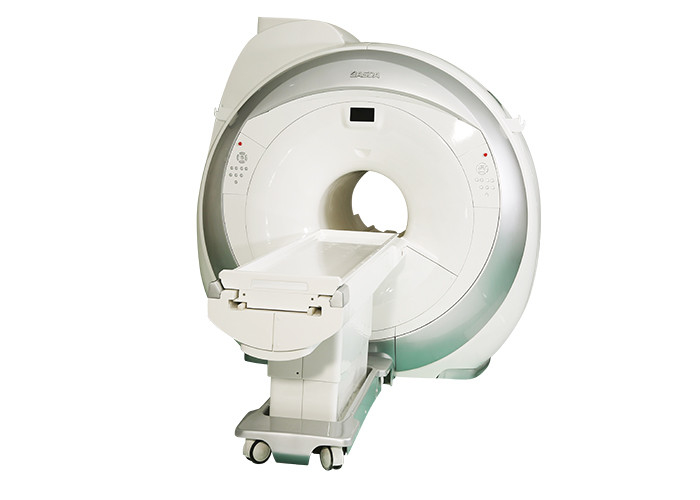
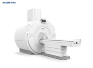
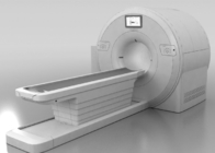
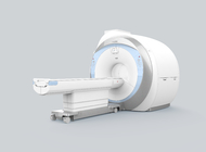
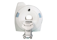
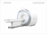
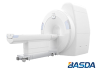
Bstar-150 Wide-bore Superconducting MRI system
Bstar-150 Wide-bore Superconducting MRI System
Software
Bstar-150 provides comprehensive scan kit and scanning sequence, with rich clinical application of the full body, with a full range of scan plan, assist the customer’s clinical diagnosis. Advanced scanning sequence are provided as well as all routine scan, the system also can take vascular imaging, includes a broad range of capabilities supporting fast, accurate diagnosis of brain conditions and injuries. From anatomical depiction to vascular 3D reconstruction.
Bstar-150 provides the power, coils and sequences for fast, intuitive, high quality spine imaging, from routine views to advanced stitching studies.
Bstar-150 provides a versatile suite of imaging capabilities for body imaging to meet the growing demands of abdominal, pelvic, and breast exams. Including MRU/MRCP/MRM imaging, and other advanced imaging sequences. Rich post-processing software can optimize images in case necessary to help in complex diseases diagnosis, excellent image quality and abundant imaging techniques.
Workflow is critical to the efficient operation of an MRI. It impacts patient satisfaction, image quality and cost effectiveness. Bstar-150 provides full preset of clinical diagnostic scan protocol, based on rich clinical experience, corresponding arrangement and optimization have been made per related clinical application features for the scan procedure and sequence parameters of the preset scan method.With unmatched clinical capability, Bstar-150 provides an unprecedented ability to acquire complete exams with a motion compensated scan technique.
By adopting the brand-new phase array coil and scan control software, application of parallel acquisition accelerating technique, improve most sequence scanning speed, on the premise of image quality guarantee, accelerating factor can reach 6 or more as the maximum. Bstar-150 system keeps pace with international design and research, keeps improving the present clinical functions to provide lifetime free software upgrade for its users as well as let the hospital enjoy the most advanced MRI clinical application techniques.
The RF system, delivers integrated coil technology to simplify patient positioning and extend coverage with high SNR, providing extremely fast scan solution, with the help of brand-new multi-channel coil and parallel accelerating technique, it gives higher spatial resolution of head images, clearer display of anatomic structure and lesions, and less artifact.
For restless patients and those who can’t cooperate, the preset extremely fast scan sequence can be used, which is very appropriate for fetus, newborns and infants.
K_Rotat artifact erasing technique, by using stripped K space rotating filling method to complete acquisition, can completely erase the effect of movement artifact on images and greatly improve SNR at the same time to provide guarantee for accurate diagnosis.
scDWI (optional)
In DWI, acquisition of low-b-value has better tissue SNR, but is not sensitive enough to water proton;
Acquisition of high-b-value has better DW sensitivity of water proton, but reduces the tissue SNR as well as the lesion location function;
By using special computation method, scDWI only needs one single DWI sequence to complete the calculation of multi-b-value within certain range, min. 100s/mm².
Meanwhile, comparing to direct acquisition of DWI images, scDWI technique greatly improved the SNR of high-b-value DW image, avoid distortion of high-b-value images, very good for lesion diagnosis.
Why Choose Basda Medical?
Employee Distribution
- A skillful, dedicated and experienced R&D team. 30%+ R&D team.
- Participated in the China's first generation of independent intellectual property rights of electromagnetic magnetic resonance
- 6% ~ 10% sales revenue is reinvested in R&D
Bstar-150 Specification.
| No. |
Item |
Specification |
| 1 |
Magnet System |
|
| 1.1 |
Magnet Type |
Superconducting |
| 1.2 |
Field Strength |
1.5T |
| 1.3 |
Shielding Method |
Active |
| 1.4 |
Shimming Method and Type |
Active + Passive + Dynamic |
| 1.5 |
Magnet stability |
≤0.1ppm/h |
| 1.6 |
Homogeneity (DSV, VRMS) |
|
| |
10cm |
≤0.01ppm |
| 1.7 |
Length of magnet(exclude cover) |
150cm |
| 1.8 |
Inner diameter of magnet |
700mm |
| 1.9 |
5 Gauss fringe field(X,Y,Z axis) |
≤2.5m x 2.5m x 4.0m |
| 1.10 |
Liquid helium boil off |
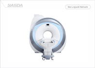
0 cc/hr |
| 1.11 |
Filling liquid helium period |
≥4 Years |
| 1.12 |
Liquid helium “zero”consumption technology |
Yes |
| 2 |
Gradient System |
|
| 2.1 |
Maximum gradient field (single axis, invalid) |
41mT/m |
| 2.2 |
Maximum gradient slew rate(single axis, invalid) |
187mT/m/ms |
| 2.3 |
Minimum gradient rise time |
0.22ms |
| 2.4 |
Maximum gradient field and slew rate reached at the same time |
Yes |
| 2.5 |
Gradient cooling system |
Water cooling |
| 2.6 |
Full digital real-time transmit and receiving gradient control system |
Yes |
| 3 |
RF System |
|
| 3.1 |
Max RF amplifier power |
18kW |
| 3.2 |
Center frequency |
63.87MHz |
| 3.3 |
Real-time digital RF energy monitoring |
Yes |
| 3.4 |
Real-time digital RF short-term accumulation monitoring |
Yes |
| 3.5 |
Real-time digital RF long-term accumulation monitoring |
Yes |
| 3.6 |
Parallel working RF receiver channels |
8/16 |
| 3.7 |
Parallel working RF A/D converters |
8/16 |
| 3.8 |
Each coil unit has corresponding pre-amplifier |
Yes |
| 3.9 |
Parallel acquisition technology platform |
Yes |
| 3.10 |
Maximum bandwidth of each parallel acquisition receive channels |
≥1.0MHz |
| 3.11 |
Transmission bandwidth |
550kHz |
| 3.12 |
Maximum receiver signal resolution |
≥16bit |
| 3.13 |
Fully digital RF system |
Yes |
| 3.14 |
RF receiving amplifier noise level |
≤0.45dB |
| 3.15 |
Multi-channel phased array receiver coil |
|
| 4 |
Scan environment |
|
| 4.1 |
Patient table driven mode |
Motor-driven |
| 4.2 |
Position accuracy |
≤0.5mm |
| 4.3 |
Table length |
2400mm |
| 4.4 |
Horizontal movement range |
2055mm |
| 4.5 |
Horizontal motion maximum speed |
≥200mm/s |
| 4.6 |
Patient table lowest height |
675mm |
| 4.7 |
Maximum patient weight |
225kg |
| 4.8 |
Feet-first entry mode |
Yes |
| 4.9 |
Table can be controlled by machine cover in case of emergency |
Yes |
| 4.10 |
Table control system on both side of rack |
Yes |
| 4.11 |
Lighting, ventilation, call system |
Be able to adjust |
| 5 |
Computer system |
|
| 5.1 |
MRI software |
BASDA |
| 5.2 |
System software |
WINDOWS 7 |
| 5.3 |
CPU |
≥3.1GHz |
| 5.4 |
Main memory |
≥4GB |
| 5.5 |
Hard disk |
≥500GB |
| 5.6 |
Color LCD monitor |
24” |
| 5.7 |
Monitor resolution |
1920×1200 |
| 5.8 |
External storage |
DVD/USB |
| 5.9 |
DICOM3.0 |
Yes |
| 6 |
Scanning parameter |
|
| 6.1 |
Spin-echo sequence |
Yes |
| 6.1.1 |
SE 2D/3D |
Yes |
| 6.1.2 |
FSE 2D/3D |
Yes |
| 6.1.3 |
FSE sharing |
Yes |
| 6.1.4 |
Single shot FSE |
Yes |
| 6.1.5 |
Spin echo fat-suppression imaging |
Yes |
| 6.1.6 |
Spin echo frequency fat suppression imaging |
Yes |
| 6.1.7 |
Spin echo water suppression imaging |
Yes |
| 6.2 |
GRE 2D/3D |
Yes |
| 6.3 |
DWI |
Yes |
| 6.3.1 |
Max. b value |
10000 |
| 6.4 |
IR sequence |
Yes |
| 6.4.1 |
IR |
Yes |
| 6.4.2 |
FIR(water / fat suppression) |
Yes |
| 6.4.3 |
FLAIR |
Yes |
| 6.4.4 |
STIR |
Yes |
| 6.4.5 |
Water-fat Separation |
Yes |
| 6.5.1 |
Pre-saturation |
Yes |
| 6.6 |
Gating |
Yes |
| 6.7 |
Accelerated sequence |
Yes |
| 6.8 |
Anti-movement scanning technology |
Yes |
| 6.9 |
Min. 2D thickness |
0.1mm |
| 6.10 |
Min. 3D thickness |
0.05mm |
| 6.11 |
Max. FOV |
50cm |
| 6.12 |
Min. FOV |
1cm |
| 7 |
Advanced imaging technology |
|
| 7.1 |
Body Imaging |
Yes |
| 7.1.1 |
Ultra-fast imaging technology |
Yes |
| 7.1.2 |
Phase / de-phase imaging technology |
Yes |
| 7.1.3 |
MR cholangiopan-creatography (MRCP) |
Yes |
| 7.1.4 |
MR urography (MRU) |
Yes |
| 7.1.5 |
MR Myelography (MRM) |
Yes |
| 7.2 |
Neuro imaging |
Yes |
| 7.3 |
Diffusion weighted imaging set |
Yes |
| 7.3.1 |
Isotropic acquisition |
Yes |
| 7.3.2 |
ADC measurement |
Yes |
| 7.4 |
MR angiography(MRA) |
Yes |
| 7.4.1 |
2D/3D TOF technology |
Yes |
| 7.4.2 |
Continuous multi-layer 3D TOF technology |
Yes |
| 7.4.3 |
Contrast enhanced MRA |
Yes |
| 7.4.4 |
Magnetization transfer (MTC) |
Yes |
| 7.4.5 |
Maximum intensity projection |
Yes |
| 7.5 |
Susceptibility weighted imaging (SWI) |
Yes |
| 7.6 |
Parallel acquisition technology |
Yes |
| 7.6.1 |
Algorithm based on image |
Yes |
| 7.6.2 |
Algorithm based on K-space |
Yes |
| 7.6.3 |
Max. parallel acquisition acceleration factor |
4 |
| 7.6.4 |
Automatic calibration technology |
Yes |
| 7.6.5 |
Applied direction of parallel acquisition factor |
X,Y,Z |
| 7.7 |
Artifact correction technology |
Yes |
| 7.7.1 |
Fluid compensation |
Yes |
| 7.7.2 |
Respiratory compensation |
Yes |
| 7.7.3 |
Head motion artifact correction |
Yes |
| 7.7.4 |
Elimination of magnetic sensitive artifact |
Yes |
| 7.7.5 |
Eddy current adaptive correction |
Yes |
| 7.7.6 |
Gradient linearity correction |
Yes |
| 7.7.7 |
Multi-echo phase correction |
Yes |
| 7.8 |
Rapid automatic correction technology |
Yes |
| 7.9 |
Automatic frequency tracking technology |
Yes |
| 7.10 |
Automatic coil recognition technology |
Yes |
| 7.11 |
Automatic phase correction technology |
Yes |
| 7.12 |
Echo navigation technology |
Yes |
| 7.13 |
RF balance drive technology |
Yes |
| 7.14 |
RF de-phase technology |
Yes |
| 7.15 |
Gradient de-phase technology |
Yes |
| 7.16 |
Section scanning technology |
Yes |
 Your message must be between 20-3,000 characters!