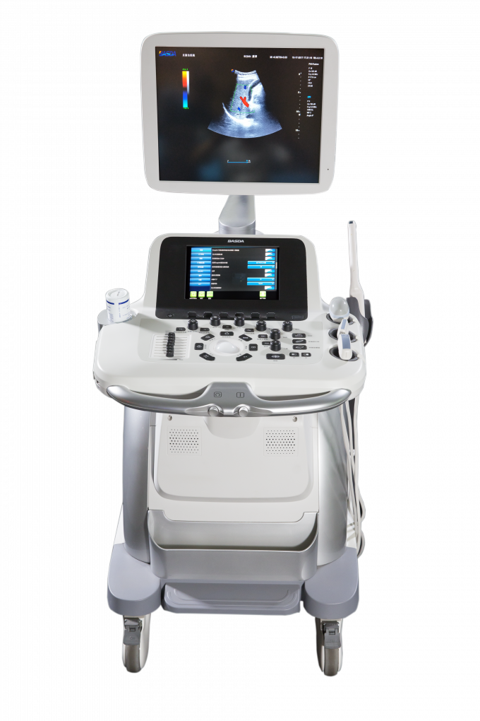
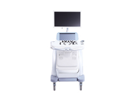
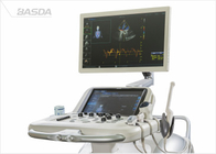
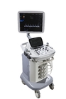
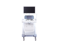
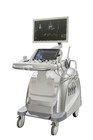
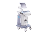
10inch Touchscreen 4D Ultrasound Equipment For Obstetrics Gynecology BTH-150S
As a leading and innovative medical technology and digital solutions provider in China, Basda Medical was established in 2000. Located in Shenzhen, the frontier city of China’s “reform and opening up”, Basda Medical has emerged and developed as a national high-tech company focusing on R&D, production, trading, and services on medical imaging, digital solutions and radiotherapy products.
The core technicalstaffs atBasda Medicalare the one of the earliest technical teams engaging in medical magnetic resonance R&D in China. Our company has obtained several hundred intellectual property rights such as patents, software copyright, proprietary technologies, etc. as the core R&D team collaborate to break through the technical barriers, master the key algorithms, conquer the main components and achieve the system innovation. Insisting on self-innovation while rooted in effective cooperation mechanisms on production, study, research and clinical trial, Basda Medical has established long-term partnership with China’s best knownuniversities such as Peking University, Harbin Institute of Technology, Southern Medical University and etc.
Basda Medicalwon numerous honors and was qualified as "National High-tech Enterprise", “Second Place of National Technology Invention", "Key Hi-tech Enterprise of State Torch Program", "First Place of China Machinery Industry Science and Technology Award", "ProvincialHigh Field MRI System Engineering and Technology R&D Center", "Shenzhen Technology Innovation Award", “Intellectual Property Management System”, etc.. Furthermore, numerous technology innovation and projects gained financial support and partnering from major investment institutions, national and regional government.
Basda Medical specializes in progressive medical imaging technologies including 9 series with over 100 products. Today, weare pioneering medical imaging trends with the MRI series, and are rapidly developing X-Ray series such as CT scanner and DR, as well as color doppler ultrasound products,and the radiotherapy products.
BTH-150S Specification
1 Application
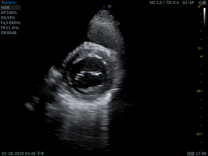
It is mainly applied for ultrasonic diagnosis and scientific research of abdomen, obstetrics and gynecology, reproduction, blood vessels, heart, superficial tissue, breast, neonates, surgery ,nerves, etc.
2 Specification:
2.1 Include:.
2.1.1 High resolution B imaging
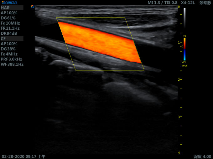
2.1.2 Color doppler imaging (CDI )
2.1.3 Pulse wave doppler (PWD)
2.1.4 Continuous wave doppler (CWD)
2.1.5 Power doppler imaging (PDI)
2.1.6 Direction Power doppler imaging (DirPDI)
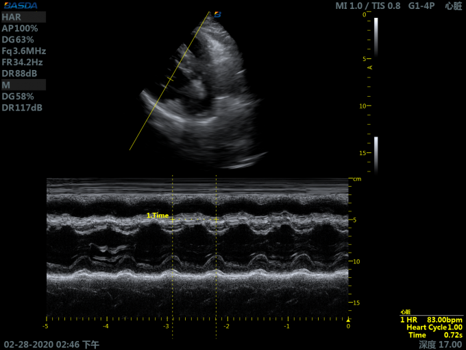
2.1.7 M mode imaging
2.1.8 Tissue doppler imaging
2.1.9 Tissue harmonic imaging
2.1.10 Harmonic fusion imaging
2.1.11 Spatial composite imaging
2.1.12 Adaptive speckle noise suppression
2.1.13 Extend imaging
2.1.14 Parallelogram imaging
2.1.15 Trapezoid imaging
2.1.16 Smart one key imaging optimization
2.1.17 Freehand 3D imaging
2.1.18 Static 3D imaging
2.1.19 Real-time 3D/4D imaging
2.1.20 Optional anatomical M imaging
2.1.21 Optional color M mode imaging
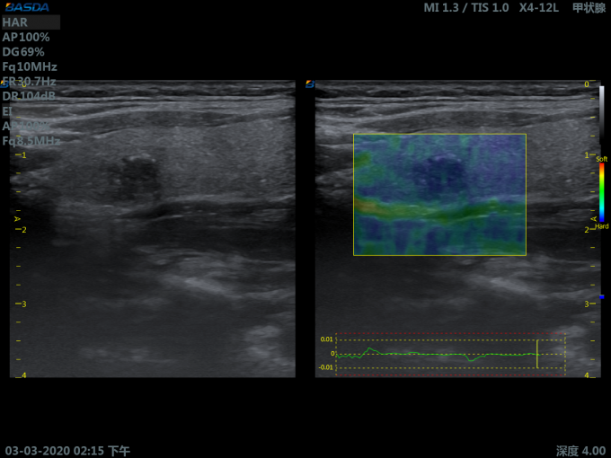
2.1.22 Optional elastography imaging
2.1.23 Optional panorama imaging
2.1.24 Optional strain rate imaging
2.2 Measurement and analysis:
2.2.1 Preset parameters: for different examined organs, the optimal image examination parameters are preset to reduce the adjustment during operation; users are permitted to define and save preset parameters
2.2.2 General measurement
2.2.3 Obstetrics measurement
2.2.4 Gynecology measurement
2.2.5 Vessel measurement
2.2.6 Urology measurement
2.2.7 Cardiac measurement
2.2.8 Abdomen measurement
2.2.9 User defined measurement
2.2.10 Doppler blood flow measurement
2.2.11 Small organs and superficial tissue measurement
2.2.12 Auto doppler spectrum tracking and analysis
2.2.13 *Auto measurement of intima-media thickness
2.3 Patient data management:
2.3.1 Patient data include: personal information, image, cine, report, etc
2.3.2 Patient data establishment, storage, playback, modification, retrieval and printing
2.3.3 Built-in integrate ultrasound workstation
2.3.4 Image storage format: PNG, JPEG, BMP
2.3.5 Cine storage format: AVI
2.3.6 Optional DICOM 3.0
3 Technical parameters:
3.1 System general configure:
3.1.1 Full digital beamformer
3.1.2 Multibeam parallel processing
3.1.3 Dynamic focusing and apodization
3.1.4 A/D sampling: ≥ 14 BIT
3.1.5 Monitor: ≥19 inch, high resolution LED color monitor that can be moved up, down, left and right
3.1.6 Touchscreen: ≥10 inch,high sensitivity capacitive touchscreen
3.1.7 Control panel: ergonomic design, lifted and rotated
3.1.8 *Built-in couplant heater
3.1.9 Transducer interfaces:≥4, fully activated, transducer can be connected to any connectors freely
3.1.10 Interfaces: network, DVI, USB
3.1.11 Built-in DVD recorder
3.1.12 Harddisk: ≥ 1T
3.1.13 Support U disk or mobile HDD storage
3.1.14 Support black white printer and color printer
3.2 Transducer specification:
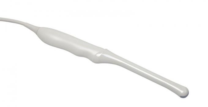
3.2.1 Support transducer type: convex array, linear array, phased array, endocavity, volume
3.2.2 Supports biopsy guide
3.2.3 Visual and adjustable transducer frequency ≥4, all modes of B ,color and doppler frequency are adjusted independently
3.2.4 Frequency range:
- Convex array transducer frequency: 2.5-5.5 MHz
- Phased array transducer frequency: 1.7-5.0 MHz
- Linear array transducer frequency: 4.0-14.0 MHz
- Endocavity transducer frequency: 3.0-10.0 MHz
- Volume transducer frequency: 2.0-6.0 MHz
3.2.5 Imaging modes:
- Convex array transducer: B/M/CDI/PWD
- Linear array transducer: B/M/CDI/PWD
- Endocavity transducer: B/M/CDI/PWD
- Phased array transducer: B/M/CDI/PWD/CWD
- Volume transducer: B/M/CDI/PWD/3D/4D
3.3 B imaging parameters:
3.3.1 Display mode: B, B/B,4B, M,B/M
3.3.2 Gain adjustment: TGC ≥8; LGC ≥8
3.3.3 Transmit beam focusing: ≥7
3.3.4 Scan lines density: ≥512
3.3.5 Gray scale ≥ 256
3.3.6 *Scanning depth ≥ 30cm
3.3.7 Visual and adjustable dynamic range: 30 ~ 180dB
3.3.8 B images playback ≥10000 frames
3.3.9 B scanning rate:
- phased array transducer: when depth is 18 cm, the full-field scanning
- frame rate ≥ 65 f/s
- convex array transducer: when depth is 18 cm, the full-field scanning
- frame rate ≥ 50 f/s
3.4 Color doppler imaging parameters:
3.4.1 Type: CDI, PDI, Dir PDI
3.4.2 Display modes: B/CDI, B/PDI, B/Dir PDI
3.4.3 B and color dual real-time contrast
3.4.4 Zero movement: ≥8 levels adjustable
3.4.5 Steer angle: linear array transducer image steer range: -20°~ +20°
3.5 Spectral doppler imaging parameters:
3.5.1 Type: PWD, CWD, HPRF
3.5.2 Display modes: B/PWD, B/CWD, B/HPRF, B/CDI/PWD, B/CDI/CWD
3.5.3 The maximum blood flow velocity : ≥5 m/s by PWD ; ≥10 m/s by CWD
3.5.4 The lowest blood flow velocity:≤0.5 mm/s (non-noise signal)
3.5.5 Sampling width : 0.5 - 30 mm
 Your message must be between 20-3,000 characters!