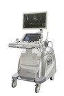
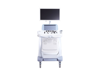
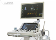
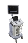
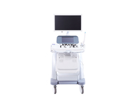
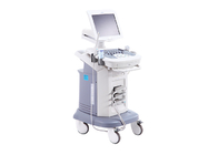
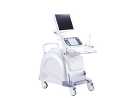
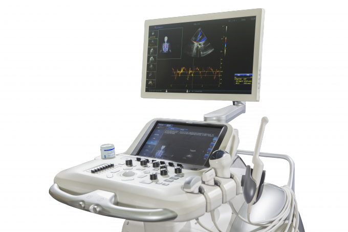
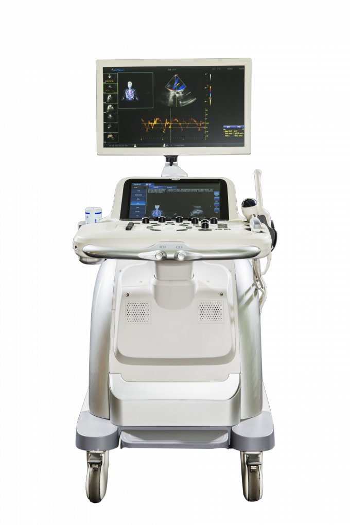
BTH-300S Color Doppler Ultrasound doppler spectrum 4 sockets 4D Large touch screen 220V/110V
Product Presentation
• Innovative RF Ultrasound Platform
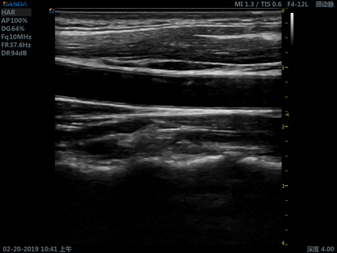
• 25MHz High Frequency System
• 128 Physical transceiver system
• 192 Array High Frequency Linear Transducer
• 21.5-inch Medical Grade LED HD Monitor
• 14-inch Hypersensitive Touch Screen
• 5 Transducer Extended Connector Types---- Abdominal, Cardiac, Intracavitary, Small Parts, 4D
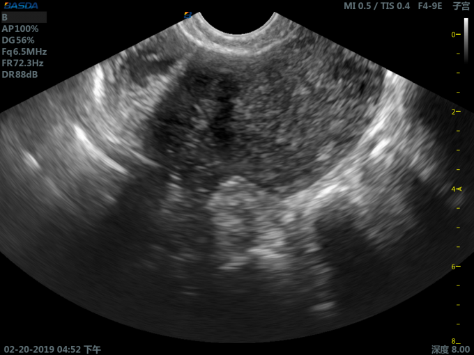
Advanced Technology for Obstetrics and Gynecology
• Outstanding Obstetrics and Gynecology
• High-Resolution 3D/4D Technology
• Amniotic Endoscopy
• Tomographic Ultrasound Imaging
• Comprehensive Gynecological Measurement Package
• Auto NT, BPD, FL, AC, HC Assessment
BTH-300S specifications
1 System overview:
1.1 Technical specifications:
1.1.1 1-25MHz High frequency system
1.1.2 Full digital broadband beam former
1.1.3 Digital high resolution B imaging
1.1.4 Color Doppler imaging
1.1.5 Power Doppler imaging
1.1.6 Direction power imaging
1.1.7 Gray-scale M Imaging
1.1.8 Color M Imaging
1.1.9 Pulsed doppler imaging
1.1.10 Continuous doppler imaging
1.1.11 Tissue doppler Imaging
1.1.12 Tissue velocity Imaging
1.1.13 Tissue velocity Imaging by M mode displaying
1.1.14 Two and three synchronization image display
1.1.15 B and color display in real time
1.1.16 Tissue harmonic imaging, reverse harmonic imaging
1.1.17 Spatial compound imaging technology
1.1.18 Speckle noise suppression technology
1.1.19 Trapezium imaging
1.1.20 B/CF/PW independent deflection
1.1.21 Grayscale inversion mode
1.1.22 One-click Automatic optimization
1.1.23 Zoom to full screen
1.1.24 Real-time local zoom
1.1.25 Focus ≥ 8
1.1.26 Dynamic range: 30dB~280dB, step by 2dB
1.1.27 High quality three dimensional / four dimensional imaging
1.1.28 Ultrasonic tomography
1.1.29 Render mode: maximum, minimum, X mode, smoothing mode, surface mode
1.1.30 Arbitrary removal of editing (Magic cut )
1.1.31 Optional volume ultrasound tomography
1.1.32 Optional panoramic imaging
1.1.33 Optional full range M
1.1.34 Optional ECG
1.1.35 Optional Elastography
1.1.36 Optional ultrasound teaching instruction function, including anatomy diagram, scanning techniques, standard ultrasound image, scan method description
1.2 37 parameter adjustment for playback image
1.2.1 2D and M Mode 15 parameters: gain, dynamic range, TGC, reversal, filtering, automatic tissue optimization, rotate, flip vertical, grayscale maps, pseudo color, frames on average, spatial smoothing, spot removal, edge enhancement, M speed
1.2.2 CFM mode 8 parameters: color auto optimization, baseline, color conversion, color map, color threshold adjustment, flash color restrain, space smooth, color M speed adjustment
1.2.3 PW Mode 14 parameters: gain, baseline, angle correction, angle fast adjustment, frequency spectrum reversed, display format, refresh rate, inhibition, gray-scale map, pseudo color, dynamic range,frequency spectrum auto optimization, automatic tracking, sensitivity tracking
1.3 Measurement and analysis
1.3.1 General measurements
1.3.2 Vessel measurement package
1.3.3 Heart measurement package
1.3.4 Gynecological measurement package
1.3.5 Urological measurement package
1.3.6 Kidney measurement package
1.3.7 Measurement and analysis of doppler blood flow
1.3.8 Obstetric measurements and fetal growth curve table
1.3.9 Intelligent three-dimensional volume measurement
1.3.10 User-defined measurement
1.3.11 Real-time tracking and measurement of doppler spectrum
1.3.12 Automatic measurement of intima-media thickness(Auto IMT)
1.3.13 Optional automatic neck-transparent layer thickness measurement (Auto NT)
1.4 Integrated image storage (film) playback to reproduce and record management components:
1.4.1 Ultrasound image static and dynamic storage, playback to reproduce the original data
1.4.2 Images can be stored as PC Compatible formats
1.4.3 Medical records management components include: patient data, reports, images, stored, modified, retrieved, and printing
1.5 Support external workstation connection
2 Technical parameters and requirements:
2.1 System General features:
2.1.1 Display: ≥ 21 inch high resolution global color LED Display, can move updown left right
2.1.2 Touch screen: ≥ 13 inch high sensitivity capacitive touch panel
2.1.3 Probe connector: ≥ 4 activated no-pin type probe connector
2.1.4 Dedicated storage tank for endocavity probe
2.1.5 Heater coupling agent
2.1.6 The operations console can be up and down, left and right
2.2 Input / Output interfaces:
2.2.1 Network interface
2.2.2 Video output: DVI , S-video
2.2.3 USB Interface: ≥ 4
2.3 Image management and recording devices:
2.3.1 Hard drive capacity: ≥ 1T
2.3.2 Integration DVD
2.3.3 Support DICOM 3.0
2.3.4 Support one key fast store to U Disk, mobile hard disk
2.3.5 Support black/white and color video printer, and quick print function
2.4 Probe specifications:
2.4.1 Probe types: convex, linear, phased array, endocavity, volume
2.4.2 Linear array probe elements ≥ 192
2.4.3 Probe frequency on display, adjust on touch-screen
2.4.4 Probe frequency ≥ 4
2.5 B image parameters:
2.5.1 Probe frequency range ( 2.0-16.0MHz )
Convex array probe 2.0-5.5MHz, harmonic frequency 4.0-6.5MHz
Phased array probe 2.0-3.5MHz, harmonic frequency 2.8-5.0MHz
Linear array probe 6.0-12.0MHz, harmonic frequency 8.0-16MHz
Endocavity probe 5.0-10.0MHz, harmonic frequency 6.5-11MHz
Volume probe 3.0-5.5MHz, harmonic frequency 4.0-6.0MHz
2.5.2 Scanning rate
Phased array probe: depth 18cm, full-field scan frame rate ≥ 100 frame / s
Convex array probe: depth 18cm, full-field scan frame rate ≥ 55 frame / s
2.5.3 Scanning line: lines density per frame ≥ 512 ultrasound line
2.5.4 Transmit beam focus: continuous focus
2.5.5 Receive: parallel process multiple signal
2.5.6 Grey scale ≥ 256
2.5.7 Digital beam forming: digital dynamic focusing, digital variable aperture and dynamic speed, A/D ≥ 14 BIT
2.5.8 Cine: gray-scale image playback ≥ 10000 picture, playback time ≥ 1000 s
2.5.9 Preconditions: preinstall best parameter for different organs
2.5.10 Gain adjustment: TGC ≥ 8
2.5.11 Side gain compensation: ≥8
2.5.12 Scan depth ≥ 36cm
2.6 Spectral doppler imaging technique parameter:
2.6.1 Support: PWD/CWD/HPRF
2.6.2 Maximum speed: PWD Blood flow velocity 9 m/s
CWD blood flow velocity 20 m/s
2.6.3 Minimum speed ≤ 1 mm/s (Non-noise signal)
2.6.4 Display mode: B, B/PWD, B/CW, B/HPRF, B/M, B/B, B/CFI/D
2.6.5 Cine: ≥ 10000 frame
2.6.6 Zero move: ≥ 12 class
2.6.7 Sample width and location range: width 0.5–10mm
2.6.8 Display control: reverse display ( left / right /up / down )
2.7 Color doppler
2.7.1 Display mode: power, velocity, three synchronization
2.7.2 Display frame rate:
Phased array probe: depth 18cm, full-field scan frame rate ≥ 24 Frame / s
Convex array probe: depth 18cm, full-field scan frame rate ≥ 15 Frame / s
2.7.3 Rotation angle: range of linear array: -20 ° ~ +20 °
2.7.4 Display control: zero move ≥ 12 levels adjustable, B compare with color in real time
2.7.5 Color enhancement: color doppler power image (CDE) , direction power doppler image(DPDI)
2.7.6 Color speed: minimum blood flow velocity ≤ 1cm /s (non-noise signal)
2.8 Ultrasonic power output adjustment for B/M, CWD, PWD, Color doppler
Our Company
Ⅰ As a leading and innovative medical technology and digital solutions provider in China, Basda Medical was established in 2000. Located in Shenzhen, the frontier city of China’s “reform and opening up”, Basda Medical has emerged and developed as a national high-tech company focusing on R&D, production, trading, and services on medical imaging, digital solutions and radiotherapy products.
Ⅱ The core technicalstaffs atBasda Medicalare the one of the earliest technical teams engaging in medical magnetic resonance R&D in China. Our company has obtained several hundred intellectual property rights such as patents, software copyright, proprietary technologies, etc. as the core R&D team collaborate to break through the technical barriers, master the key algorithms, conquer the main components and achieve the system innovation. Insisting on self-innovation while rooted in effective cooperation mechanisms on production, study, research and clinical trial, Basda Medical has established long-term partnership with China’s best knownuniversities such as Peking University, Harbin Institute of Technology, Southern Medical University and etc.
Ⅲ Basda Medicalwon numerous honors and was qualified as "National High-tech Enterprise", “Second Place of National Technology Invention", "Key Hi-tech Enterprise of State Torch Program", "First Place of China Machinery Industry Science and Technology Award", "ProvincialHigh Field MRI System Engineering and Technology R&D Center", "Shenzhen Technology Innovation Award", “Intellectual Property Management System”, etc.. Furthermore, numerous technology innovation and projects gained financial support and partnering from major investment institutions, national and regional government.
Ⅳ Basda Medical specializes in progressive medical imaging technologies including 9 series with over 100 products. Today, weare pioneering medical imaging trends with the MRI series, and are rapidly developing X-Ray series such as CT scanner and DR, as well as color doppler ultrasound products,and the radiotherapy products.
Ⅴ Specifically, our products in the permanent MRI system series have varying magnetic field strengths of 0.20T, 0.30T, 0.35T, 0.42T and 0.50T respectively. Our superconductive MRI systems have magnet filed strength range from 0.5T, 0.52T, 0.7T, 1.2T, 1.49T, 1.5T to 3.0T. On the other hand, our CT scannerseries has products with various slide widths of 16 slice, 32slice and 64 slice. Moreover, the products in the DR series include ceiling suspension DR, U-arm DR, mobile DR, digital X-Ray, DRF system, and digital mammography. Furthermore, our color doppler ultrasound system products range from portable, wireless to trolley products. Lastly, we also provide medical linear acceleratorradiotherapy series and nuclear medicine series such as positron emission computed tomography(PET-CT), single-photon emission computed tomography (SPECT), wide FOV gamma camera, etc.
 Your message must be between 20-3,000 characters!