Basda Medical continues to deliver on "technology safeguards our lives" promise as we strive to become the world’s most admired leading enterprise in the new medical imaging era. We are passionate about technology and driven by innovation, and always dedicate to excellent products and service for customers. We dream, we dare and we strive to create an effortless and healthy life for everyone.
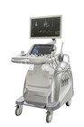
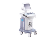
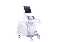
BTH-200S Specification
1 Application
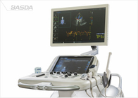
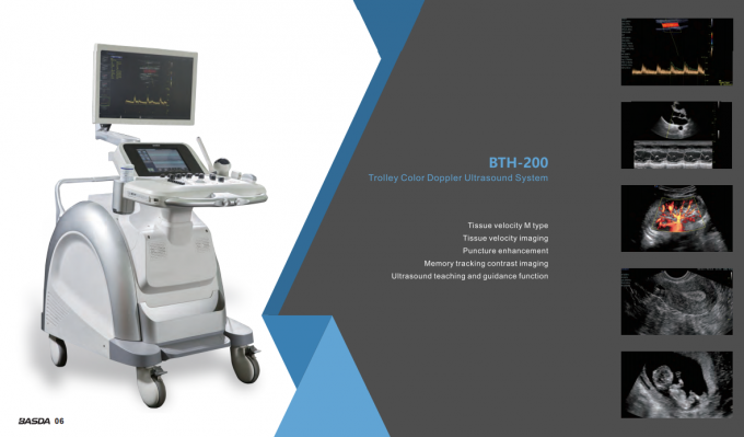
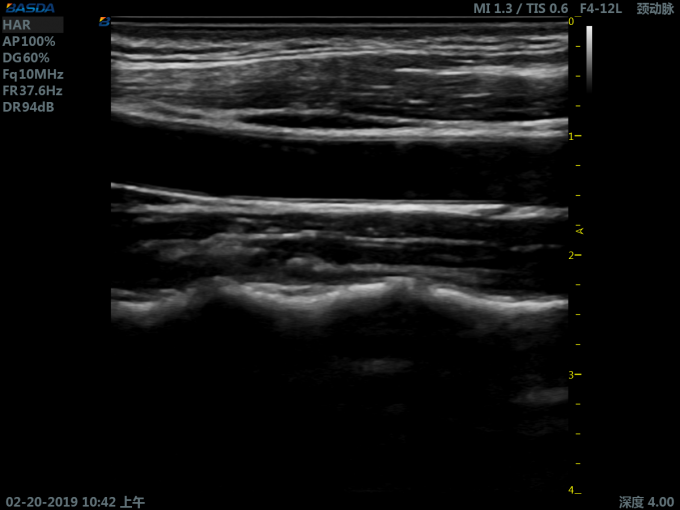
It is mainly applied for ultrasonic diagnosis and scientific research of abdomen, obstetrics and gynecology, reproduction, blood vessels, heart, superficial tissue, breast, neonates, surgery, nerves, etc
2 Specification:
2.1 Include:
2.1.1 High resolution B imaging
2.1.2 Color doppler imaging (CDI )
2.1.3 Pulse wave doppler (PWD)
2.1.4 Continuous wave doppler (CWD)
2.1.5 Power doppler imaging (PDI)
2.1.6 Direction Power doppler imaging (DirPDI)
2.1.7 M mode imaging
2.1.8 Tissue doppler imaging
2.1.9 Tissue harmonic imaging
2.1.10 Pulse reverse harmonic imaging
2.1.11 Spatial composite imaging
2.1.12 Speckle noise suppression
2.1.13 Trapezoid imaging
2.1.14 Zoom in full screen
2.1.15 Real-time zoom
2.1.16 Smart one key imaging optimization
2.1.17 Static 3D imaging
2.1.18 Real-time 3D/4D imaging
2.1.19 Ultrasound tomography imaging
2.1.20 Magic cut: edit arbitrarily for 3D imaging
2.1.21 Render types:maximum, minimum, X ray display, surface, smoothness
2.1.22 Optional volume ultrasound tomography imaging
2.1.23 Optional multi angle M imaging
2.1.24 Optional color M mode imaging
2.1.25 Optional panorama imaging
2.1.26 Optional elastography imaging
2.1.27 Optional tissue velocity imaging
2.1.28 Optional tissue velocity M imaging
2.1.29 Optional gray-scale inversion imaging
2.1.30 Optional ultrasound learning and guide function: anatomical schematic diagram, scanning manipulation diagram, standard ultrasonic image, scanning method description, etc
2.2 *37 parameters adjustment when playback images:
2.2.1 Preset parameters: for different examined organs, the optimal image examination parameters are preset to reduce the adjustment during operation; users are permitted to define and save preset parameters
2.2.2 15 parameters on B and M mode: gain, dynamic range, TGC, image rollover, gray filter, tissue optimization, image rotation, left and right rollover, gray map, pseudocolor, frame average, spatial smooth, speckle rejection, edge enhance, M speed
2.2.3 8 parameters on CDI mode: color optimization, base line, color rollover, color map,color threshold adjustment, glitter rejection, spatial smooth, color M speed
2.2.4 14 parameters on PWD mode: gain, base line, angle revise,quick angle adjustment, spectrum rollover, display format, refresh rate, rejection, gray map, pseudocolor, dynamic range, spectrum optimization, auto tracking, sensitivity tracking
2.3 Measurement and analysis:
2.3.1 General measurement
2.3.2 Obstetrics measurement
2.3.3 Gynecology measurement
2.3.4 Vessel measurement
2.3.5 Urology measurement
2.3.6 Cardiac measurement
2.3.7 Abdomen measurement
2.3.8 User defined measurement
2.3.9 Doppler blood flow measurement
2.3.10 Small organs and superficial tissue measurement
2.3.11 Intelligent volume calculation
2.3.12 Auto doppler spectrum tracking and analysis
2.3.13 Optional auto measurement of intima-media thickness(Auto IMT)
2.3.14 Optional auto neck transparent layer thickness measurement(Auto NT)
2.4 Patient data management:
2.4.1 Patient data include: personal information, image, cine, report, etc
2.4.2 Patient data establishment, storage, playback, modification, retrieval and printing
2.4.3 Built-in integrate ultrasound workstation
2.4.4 Image storage format: PNG, JPEG, BMP, TIFF
2.4.5 Cine storage format: AVI, VRD
2.4.6 Optional DICOM 3.0
3 Technical parameters:
3.1 System general configure:
3.1.1 1-25MHz high frequency system platform
3.1.2 Full digital uwb beamformer
3.1.3 A/D sampling ≥ 14 BIT
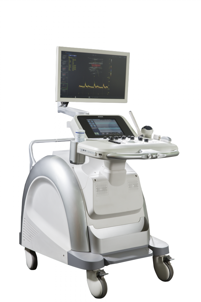
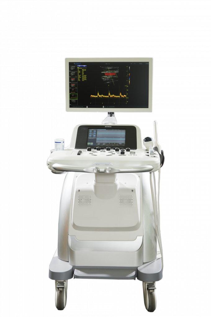
3.1.4 Monitor: ≥21 inch, high resolution LED color monitor that can be moved up, down, left and right
3.1.5 Touchscreen: ≥10 inch, high sensitivity capacitive touchscreen
3.1.6 Control panel: ergonomic design, lifted and rotated
3.1.7 *Built-in couplant heater
3.1.8 Transducer interfaces:≥3, fully activated, transducer can be connected to any connectors freely
3.1.9 Interfaces: network, DVI, USB, S-video
3.1.10 Built-in DVD recorder
3.1.11 Harddisk: ≥ 1T
3.1.12 Support U disk or mobile HDD storage
3.1.13 Support black white printer and color printer
3.2 Transducer specification:
3.2.1 Support transducer type: convex array, linear array, phased array, endocavity, volume
3.2.2 Supports biopsy guide
3.2.3 Visual and adjustable transducer frequency ≥4, all modes of B ,color and doppler frequency are adjusted independently
3.2.4 Frequency range:
Convex array transducer frequency: 2.5-6.5 MHz
Phased array transducer frequency: 2.0-5.0 MHz
Linear array transducer frequency: 6.0-16.0 MHz
Endocavity transducer frequency: 5.0-11.0 MHz
Volume transducer frequency:3.0-6.0 MHz
3.2.5 Imaging modes:
Convex array transducer: B/M/CDI/PWD
Linear array transducer: B/M/CDI/PWD
Endocavity transducer: B/M/CDI/PWD
Phased array transducer: B/M/CDI/PWD/CWD
Volume transducer: B/M/CDI/PWD/3D/4D
3.3 B imaging parameters:
3.3.1 Display mode: B, B/B,4B, M,B/M
3.3.2 Gain adjustment: TGC ≥8; LGC ≥8
3.3.3 Transmit beam focusing: ≥7
3.3.4 Scan lines density: ≥512
3.3.5 Gray scale: ≥ 256
3.3.6 Focus numbers ≥8
3.3.7 *Scanning depth ≥ 36cm
3.3.8 *Visual and adjustable dynamic range: 30 ~ 280dB, step 2
3.3.9 B images playback: ≥10000 frames, playback time: ≥1000 s
3.3.10 *B scanning rate:
phased array transducer: when depth is 18 cm, the full-field scanning
frame rate ≥ 100 f/s
convex array transducer: when depth is 18 cm, the full-field scanning
frame rate ≥ 55 f/s
3.4 Color doppler imaging parameters:
3.4.1 Type: CDI, PDI, Dir PDI
3.4.2 Display modes: B/CDI, B/PDI, B/Dir PDI
3.4.3 B and color dual real-time contrast
3.4.4 Zero movement: ≥ 12 levels adjustable
3.4.5 Steer angle: linear array transducer image steer range: -20°~ +20°
3.4.6 Color scanning rate:
phased array transducer: when depth is 18 cm, the full-field scanning frame rate ≥ 24 f/s
convex array transducer: when depth is 18 cm, the full-field scanning frame rate ≥ 15 f/s
3.5 Spectral doppler imaging parameters:
3.5.1 Type: PWD, CWD, HPRF
3.5.2 Display modes: B/PWD, B/CWD, B/HPRF, B/CDI/PWD, B/CDI/CWD
3.5.3 The maximum blood flow velocity : ≥10 m/s by PWD ; ≥20 m/s by CWD
3.5.4 The lowest blood flow velocity:≤1 mm/s (non-noise signal)
3.5.5 Spectral images playback: ≥10000 frames
3.5.6 Zero movement ≥12 levels
3.5.7 Sampling width : 0.5 - 28 mm
3.5.8 B/CDI/PWD steer independently
3.6 Ultrasound power output adjust for B, M, PWD, CWD,CDI
Reasons to Choose Basda
1. China’s "National High-tech Enterprise"
2. China’s most MRI registration certificates company with manufacturing history of more than 20 years
3. Topped the best-selling MRI in China for consecutive 6 years
4. Known as the one of the two earliest manufacturers of 3.0T MRI system in China
5. Successfully developed and commercialized the 2nd specialized extremity 0.2T Permanent MRI which expanded the company product line from extremity to whole body, from human to veterinary
6. Successfully developed and commercialized the first 1.2T Superconductive MRI
 Your message must be between 20-3,000 characters!